Home
/ Compact Bone Diagram Labeled : Compact Bone Labeling : Production of compact bone c.
Compact Bone Diagram Labeled : Compact Bone Labeling : Production of compact bone c.
Compact Bone Diagram Labeled : Compact Bone Labeling : Production of compact bone c.. There are small canals that run through the bone, which allow blood vessels to penetrate it. Bones have numerous small gaps between their cells and the components of the extracellular matrix (among others, collagen fibers, water, electrolytes, and glycosaminoglycans). Online quiz to learn structure of compact bone; Compact bone is the denser, stronger of the two types of bone tissue (figure 6.12). A diagram of the anatomy of a bone, showing the compact bone.
Start studying compact bone labeling. Online quiz to learn structure of compact bone; Bodytomy provides a labeled diagram of the haversian system to help you understand its structure and function. Compact bone is formed in concentric circles. Bone marrow diagram, compact bone diagram quiz, compact bone slide labeled, diagram long bone, labeled compact bone model, human anatomy, bone marrow diagram, compact bone diagram quiz, compact bone slide labeled, diagram long bone, labeled compact bone model.
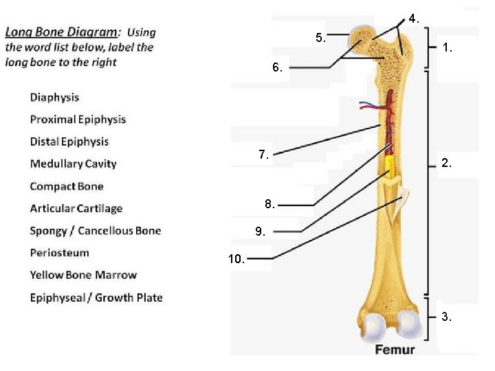
Holes Essentials Of Human Anatomy Physiology Unit 3 from slidetodoc.com Add to favorites 25 favs. Label the structural components of bone tissue in the diagram: Spongy bone is used for more active functions of the bones, including blood cell production and ion exchange. The shaft is composed of compact bone this page is about compact bone labeled diagram,contains anatomy & physiology i bis 240: As we have learned, bone consists of periosteum, compact bone, spongy bone, endosteum, and bone marrow. Learn vocabulary, terms, and more with flashcards, games, and other study tools. Human bone generally comprises osseous tissue, an outer coating called a periosteum, and bone marrow. The shafts found in long bones are also compact bones.
90 introduction to the skeletal system exercise 7
Compact bone histology slide structure with diagram. I am not an expert on this subject, so i was wondering if anyone could put their input on i don't like way you've shown the cartilage. A diagram of the anatomy of a bone, showing the compact bone. Add to favorites 0 favs. Human bone generally comprises osseous tissue, an outer coating called a periosteum, and bone marrow. Under magnification you can clearly see the system of concentric circles that forms compact bone. The remainder is cancellous bone, which has a spongelike appearance with numerous large spaces and is found in the. Good, here in this part, i am going to describe the structure of compact bone. The two main structural components typically include spongy bone on the interior, with an outer layer of compact bone. Do you want to learn the details of the histology of compact bone with labelled diagram and authentic slide images? Label the structural components of bone tissue in the diagram: The smallest units of bones are found inside the compact bone. It can be found under the periosteum and in the diaphyses of long bones, where it provides support and protection.
Long bone diagram labled / bone anatomy diagrams for coloring and labeling with reference and summary : Compact bone histology slide structure with diagram. 1278 x 720 jpeg 92 кб. Add to favorites 25 favs. Compact and spongy.the names imply that the two types differ in density, or how tightly the tissue is packed together.
Cartilage And Bones Anatomy Bones Structure Of Bone Medical from i.pinimg.com Structure and parts of long bones. A diagram of the anatomy of a bone, showing the compact bone. Due to the strong nature of compact bone, compared to spongy bone, it is the preferred tissue for strength. You need to get 100% to score the 10 points available. However, compact bones also serve a function in storing and releasing calcium to the. It can be found under the periosteum and in the diaphyses of long bones, where it provides support and protection. Label the structural components of bone tissue in the diagram: The compact bone is the main structure in the body for support, protection, and movement.
In these labeled examples, a human femur is represented without identifying many of the unique characteristics that help differentiate the femur bone from other.
There are two types of bone tissue: Bone model label the parts of a compact bone. Human bone generally comprises osseous tissue, an outer coating called a periosteum, and bone marrow. It can be found under the periosteum and in the diaphyses of long bones, where it provides support and protection. You can think of compact bone as being very similar. Production of compact bone c. The smallest units of bones are found inside the compact bone. The cells of compact bone, which is also called cortical bone, appear to be tightly packed into a solid mass. Add to favorites 25 favs. On this page, you will find two images i created that illustrate the parts of a long bone and long bone structure. Compact bone is formed in concentric circles. Bodytomy provides a labeled diagram of the haversian system to help you understand its structure and function. The shaft is composed of compact bone this page is about compact bone labeled diagram,contains anatomy & physiology i bis 240:
In long bones, as you move from the outer cortical compact bone to the inner medullary cavity, the bone transitions to spongy bone. Compact bone labeled diagram (page 1) bone histology, general overview. Add to favorites 0 favs. There are small canals that run through the bone, which allow blood vessels to penetrate it. Structure and parts of long bones.
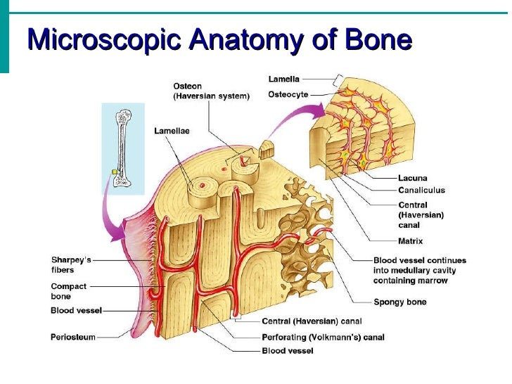
32 Label Compact Bone Labels Design Ideas 2020 from image.slidesharecdn.com The diagram above shows a longitudinal view of an osteon. Good, here in this part, i am going to describe the structure of compact bone. Compact bone is the denser, stronger of the two types of osseous tissue (figure 6.3.6). Bodytomy provides a labeled diagram of the haversian system to help you understand its structure and function. On this page, you will find two images i created that illustrate the parts of a long bone and long bone structure. Bones have numerous small gaps between their cells and the components of the extracellular matrix (among others, collagen fibers, water, electrolytes, and glycosaminoglycans). In long bones, as you move from the outer cortical compact bone to the inner medullary cavity, the bone transitions to spongy bone. Online quiz to learn structure of compact bone;
The two main structural components typically include spongy bone on the interior, with an outer layer of compact bone.
The diagram above shows a longitudinal view of an osteon. 90 introduction to the skeletal system exercise 7 Under magnification you can clearly see the system of concentric circles that forms compact bone. Online quiz to learn compact bone diagram; Some, mostly older, compact bone is remodelled to form these haversian systems (or osteons). Due to the strong nature of compact bone, compared to spongy bone, it is the preferred tissue for strength. I am not an expert on this subject, so i was wondering if anyone could put their input on i don't like way you've shown the cartilage. A diagram of the anatomy of a bone, showing the compact bone. There are small canals that run through the bone, which allow blood vessels to penetrate it. Labeling portions of a long bone. In these labeled examples, a human femur is represented without identifying many of the unique characteristics that help differentiate the femur bone from other. The compact bone is the main structure in the body for support, protection, and movement. Start studying compact bone labeling.